3D Printed Silicone Implant Combines Brain Cooling and Neural Recording
- Published
- Author
- Aura Moreno
- Reads
- 529
A soft 3D printed implant cooled targeted brain tissue and recorded neural activity, reducing seizure-like signals in rats during short tests.
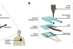
A soft implant that cools the brain while recording its electrical activity could give researchers a way to study focal cooling as a treatment for drug-resistant epilepsy. In tests on anesthetized rats, cooling reduced seizure-like brain signals within seconds. The device remains an experimental system: its external driver is large, and the animal results do not establish how it would perform in people.

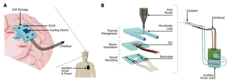
Researchers from the Leibniz Institute for Polymer Research Dresden, TUD Dresden University of Technology, the University of Sheffield and the University of Limerick built the device from printed silicone modules. It brings three functions into one implant placed on the brain’s surface: electrical recording, thermoelectric cooling and fluid circulation to remove heat.
A layered device printed from silicone
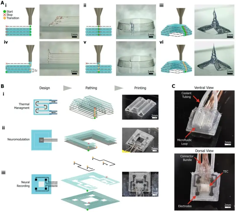
The implant measures 10 x 10 x 3 mm. The team made it using direct ink writing, extruding DOWSIL SE1700 silicone from Dow on a RegenHU 3D Discovery printer. The researchers say the process uses non-cleanroom tools and commercially available components, an approach that could lower development costs for implants combining several functions.
Three printed modules form the device. Four electrodes, made from a graphite and silicone composite, sit in the bottom layer and record electrical activity. A miniature thermoelectric cooler (TEC) occupies the middle layer. When current passes through it, the TEC transfers heat from one face to the other. The top layer contains a water channel that carries heat away from the TEC’s warm side.
Each silicone layer was printed in one continuous path, avoiding seams that could weaken the structure. The team then cured the stacked modules together to make a single implant. The design matters because cooling a patch of brain tissue also means managing the heat displaced by the cooler. The integrated water path addresses that heat load within the same soft structure as the electrodes and TEC.
Cooling performance and heat management
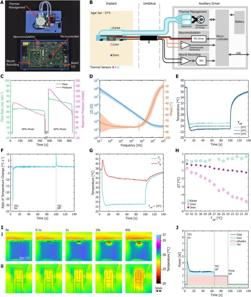
During testing, a separate driver unit powered the implant, circulated water and adjusted current to the TEC using temperature readings. The unit measures 17 x 8 x 5 cm. The design goal was to prevent surrounding brain tissue from warming by more than 1°C as heat moved through the cooler.
Modeling indicated that natural cerebrospinal fluid flow would not keep the TEC’s warm side within that temperature limit. The team therefore used forced water circulation. In agar gel held at 37°C to mimic brain tissue, the system reached 23°C in about 5 seconds and maintained its target temperature within ±0.25°C. Testing showed that heat from the warm side remained inside the implant, while the cooling effect extended 1 to 3 mm below it. Steady cooling drew 2.6 W.
Those measurements have a clear limitation: agar does not reproduce blood flow, which can carry heat through living tissue. The researchers say the gel test likely overstates how far cooling would spread in brain tissue. The reported depth should therefore be read as a result under the test conditions, rather than a direct measure of performance in people.
Short cooling events reduced seizure-like signals
The team tested the implant in nine anesthetized rats. Researchers induced seizure-like activity using 4-aminopyridine, delivered through a NeuroNexus depth probe that also recorded activity inside the cortex. The surface implant then applied 60-second cooling events, with settings ranging from 25°C to 15°C.
At every tested temperature, cooling lowered average brain signal energy, and the signals weakened within seconds of activation. The 15°C setting was slightly less effective than the milder settings. The researchers caution that this unexpected pattern needs more study: it differs from prior work and was observed in a small number of trials.
The implant’s own electrodes captured the most consistent reductions in the delta and beta brainwave bands. At 18°C, delta-band signal energy fell by 48 percent ± 35 percent. Activity returned tens of seconds after cooling stopped. In these experiments, then, cooling temporarily dampened seizure-like activity; it did not end it.
What remains before longer-term use
The measured cooling depth of 1 to 3 mm is similar to the thickness of the human neocortex, according to the researchers. That makes the design a possible fit for seizure sources in that layer. A deeper or more widely distributed source could call for multiple cooling contacts or a different implant shape.
Long-term implantation would also require substantial changes to the support hardware. The current driver has a volume of 680 cm³; the authors say it would need to approach the 10 to 20 cm³ volume of chest-implanted pacemakers. Other requirements include a reliable connector, an integrated power supply, a sealed titanium housing and aging tests. Wireless power transfer is one route the team identifies for powering a future system.
Recording could eventually help control the cooling. The authors suggest that neural signals might trigger cooling automatically, drawing on adaptive deep brain stimulation for Parkinson’s disease, where output can respond to beta-band activity. That is a proposed direction, not a capability demonstrated by this implant.
The work extends earlier printed implant research that focused on electrical recording or stimulation. A 2020 study co-authored by Ivan R. Minev described soft printed implants that stimulated the spinal cords of injured rats and zebrafish. The new device adds temperature control and heat removal to the same kind of integrated approach. The study, “Printing-Assisted Integration of Thermal, Fluidic and Electrical Components in Implants for Focal Brain Cooling,” was conducted by Spencer Ryan Moore, Naomi King, Aruã Clayton Da Silva, Clare Howarth, Jason Berwick, Thomas E. Paterson and Ivan R. Minev. Funding came in part from the European Research Council, with support from the Sheffield University Neuroscience Institute.