VINERA Surgical Imaging Platform Combines 2-Megapixel Sensors with 100-Micron Illumination Fiber
- Published
- Author
- Semiconductor For You
- Reads
- 993
OMNIVISION and MFI combine medical imaging, processing and a 100 µm laser fiber in VINERA for compact endoscopic and robotic systems.
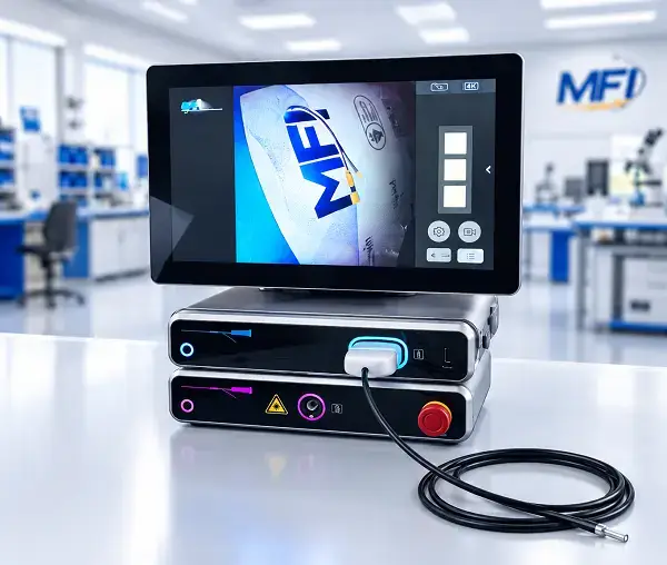
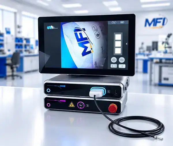

VINERA brings image capture, processing, patient isolation and illumination into one surgical visualization platform aimed at small-diameter endoscopes and robotic instruments. Announced by OMNIVISION and Myriad Fiber Imaging (MFI) on November 16, 2026, the joint development pairs OMNIVISION medical imaging components with MFI’s system integration and a laser illumination fiber as small as 100 microns (µm).
The engineering problem is at the distal end of the instrument, where the imaging and lighting hardware must fit within limited space. Designers need to balance image resolution and quality against illumination, thermal management and the scope’s diameter. The companies position VINERA as a way for medical-device manufacturers to work with an integrated set of technologies rather than build each part of the imaging system independently.
Imaging and processing components
The platform includes OMNIVISION’s OVMed® medical image sensors and image-processing technology. The named sensor options are the OCH2B10 CameraCubeChip® and OCH2B10 cable module, with resolution up to 2 megapixels (MP). The listed processing component is the OH0131 OVMed® image signal processor.
Those components provide the image-capture and processing side of the system. MFI contributes its patient-isolated post-processing architecture and integrates the components into VINERA. The announcement describes this as a platform for OEM development; it does not specify sensor dimensions, frame rates, image formats, power consumption or thermal performance. Those details would be needed to assess a particular design’s resolution, latency, electrical and packaging constraints.
The integration approach addresses a practical part of early device development: getting imaging, processing, isolation and illumination to function together in a compact assembly. OEMs still need to determine how a platform fits their instrument geometry and application requirements, but starting from a combined system could reduce the amount of custom development needed to produce a working prototype.
Illumination through a 100 µm fiber
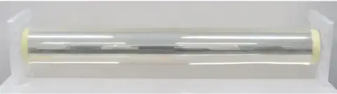
MFI’s new laser illumination fiber is specified to deliver high-intensity, wide-angle illumination through a fiber as small as 100 µm. MFI gives the light distribution as 140 degrees. The small fiber is intended to help address the space consumed by lighting in narrow endoscopes, where illumination must reach the viewing area without requiring a proportional increase in the instrument’s distal diameter.
That packaging goal involves several coupled design choices. A smaller illumination path can free space at the instrument tip, while the sensor and associated electronics also have to fit and manage heat. The companies say the combined platform is intended to support higher image quality and stronger illumination within constrained distal spaces. They have not provided measured illumination output, test conditions or thermal data, so the stated capabilities should be read as design aims rather than comparative performance results.
Tyler Gauvin, MFI president, said the fiber’s wide distribution and small size address a limitation in small-diameter endoscopes. Tehzeeb Gunja, OMNIVISION’s director of medical marketing, described the combined imaging, processing, isolation and lighting as a way to reduce integration complexity and speed development from concept to functional prototype. Both comments reflect the companies’ intended use of the platform; no prototype schedule or development-time figures were given.
Intended device applications
The companies list laparoscopy, hysteroscopy, endoluminal robotic surgery, urology and ureteroscopy among the possible applications. The platform is also aimed at flexible and rigid endoscopy, single-use and reusable endoscopes, and other small-diameter surgical visualization systems that call for high image quality and high-intensity illumination.
These applications span different instrument types and use conditions, so the listed components alone do not establish compatibility with a specific device. Manufacturers will need to evaluate the sensor, processor, illumination fiber and patient-isolation architecture against their own optical layout, packaging and system requirements. The announcement does not give pricing, availability or a finished device specification.
OMNIVISION and MFI plan to demonstrate the joint solution at OMNIVISION’s booth 8a|D01 during COMPAMED, held November 16–19, 2026, at Messe Düsseldorf in Germany. For engineers assessing compact visualization systems, the demonstration should clarify how the components are brought together; detailed performance and integration data will determine how the platform fits a given instrument design.